Turbellarians image

Aphanostoma pisae
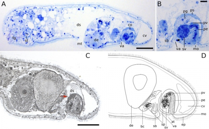
Description Sagittal sections of mature animals, anterior to the left, dorsal up. (?B) Stained after Richardson et al. (1960). (C) Stained after Heidenhain (Romeis, 1968). Red arrow marks the opening of the bursa to the digestive syncytium. (D) Schematic drawing, based on sections of several individuals. at atrium, bc bursal cap, co copulatory apparatus, cv chordoid vacuole, de developing egg, ds digestive syncytium, ep ciliated epidermis, fg frontal glands, mo male genital opening, mt mouth, pe penis, pg prostatic glands, ps penis sheath, pv prostatic vesicle, sb seminal bursa, sp sperm, st statocyst, sv seminal vesicle, va vagina. Scale bars: 50 ?m in (A, C), 10 ?m in (B)
JPG file - 129.63 kB - 800 x 496 pixels
Extra information
Original Width: 961 pixels
Original Height: 596 pixels
added on 2017-04-071 106 viewsTurbellarians taxaScan of photo Aphanostoma pisae Zauchner, Salvenmoser & Egger, 2015 accepted as Praeconvoluta pisae (Zauchner, Salvenmoser & Egger, 2015)checked Tyler, Seth 2017-04-07
Original Width: 961 pixels
Original Height: 596 pixels
 Comment (0)
Disclaimer: Turbellarians does not exercise any editorial control over the information displayed here. However, if you come across any misidentifications, spelling mistakes or low quality pictures, your comments would be very much appreciated. You can reach us by emailing info@marinespecies.org, we will correct the information or remove the image from the website when necessary or in case of doubt.