Foraminifera image

Pseudedomia persica Rahaghi, 1989
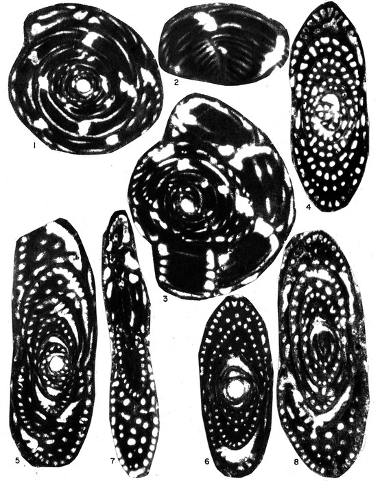
Description 1. Horizontal section showing regular whorls, preseptal canals, central thickening and a large proloculus.2. A part of subhorizontal section showing preseptal canals, sutures and subepidermal lamellae.
3. Holotype: horizontal section showing much developed last whorle and preseptal canal.
4. Subvertical section showing nearly a biumbilicate test.
5, 6. Vertical sections showing the shape of the test, peripheral chamberlets, supplimentary chamberlets in the central thickening.
7, 8. Oblique sections showing the biumbilicate test and peripheral chamberlets. JPG file - 2.60 MB - 4 097 x 5 178 pixels added on 2023-12-19101 viewsForaminifera taxaScan of photo Pseudedomia persica Rahaghi, 1989 †checked Consorti, Lorenzo 2023-12-19
 Comment (0)
Disclaimer: Foraminifera does not exercise any editorial control over the information displayed here. However, if you come across any misidentifications, spelling mistakes or low quality pictures, your comments would be very much appreciated. You can reach us by emailing info@marinespecies.org, we will correct the information or remove the image from the website when necessary or in case of doubt.