WoRMS taxon details
Phalacroma mitra F.Schütt, 1895
232491 (urn:lsid:marinespecies.org:taxname:232491)
accepted
Species
Dinophysis mitra (F.Schütt) T.H.Abé, 1967 · unaccepted
Dinophysis mitra (F.Schütt) Balech, 1967 · unaccepted (synonym)
Phalacroma dolichopterygium Jörgensen, 1923 · unaccepted (synonym)
Prodinophysis mitra (Schütt) Balech · unaccepted (synonym)
marine, terrestrial
(of Dinophysis mitra (F.Schütt) T.H.Abé, 1967) Schütt, F. (1895). Ergebnisse der Plankton-Expedition der Humboldt-Stiftung. Bd. IV. Peridineen der Plankton-Expedition. Lipsius & Tischer, Kiel. [details]
Type locality contained in Atlantic Ocean
type locality contained in Atlantic Ocean [from synonym] [view taxon] [details]
LSID urn:lsid:algaebase.org:taxname:42849
Description Cells large, broad and wedgeshaped. 70–95 μm in length and 58–70 μm in dorso-ventral width. The ventral hypothecal...
Distribution Widely distributed in warm temperate to tropical waters.
LSID urn:lsid:algaebase.org:taxname:42849 [details]
Description Cells large, broad and wedgeshaped. 70–95 μm in length and 58–70 μm in dorso-ventral width. The ventral hypothecal...
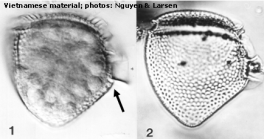
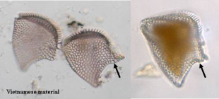
Description Cells large, broad and wedgeshaped. 70–95 μm in length and 58–70 μm in dorso-ventral width. The ventral hypothecal margin is distinctly concave below the left sulcal list. The LSL is relatively short, only half of the total cell length. Cells are widest at the base of the second rib of the LSL (left sulcal list). Thecae thick and coarsely areolated. Areolae large, some with a small central pore. [details]
Distribution Widely distributed in warm temperate to tropical waters.
Distribution Widely distributed in warm temperate to tropical waters.
[details]
[details]
Guiry, M.D. & Guiry, G.M. (2024). AlgaeBase. World-wide electronic publication, National University of Ireland, Galway (taxonomic information republished from AlgaeBase with permission of M.D. Guiry). Phalacroma mitra F.Schütt, 1895. Accessed through: World Register of Marine Species at: https://www.marinespecies.org/aphia.php?p=taxdetails&id=232491 on 2024-04-23
Date
action
by
2006-07-24 10:32:07Z
created
Camba Reu, Cibran
2007-10-17 15:07:21Z
changed
db_admin
Copyright notice: the information originating from AlgaeBase may not be downloaded or replicated by any means, without the written permission of the copyright owner (generally AlgaeBase). Fair usage of data in scientific publications is permitted.
original description
(of Dinophysis mitra (F.Schütt) T.H.Abé, 1967) Schütt, F. (1895). Ergebnisse der Plankton-Expedition der Humboldt-Stiftung. Bd. IV. Peridineen der Plankton-Expedition. Lipsius & Tischer, Kiel. [details]
basis of record Gómez, F. (2005). A list of free-living dinoflagellate species in the world's oceans. <em>Acta Bot. Croat.</em> 64(1): 129-212. [details]
additional source Steidinger, K. A., M. A. Faust, and D. U. Hernández-Becerril. 2009. Dinoflagellates (Dinoflagellata) of the Gulf of Mexico, Pp. 131–154 in Felder, D.L. and D.K. Camp (eds.), Gulf of Mexico–Origins, Waters, and Biota. Biodiversity. Texas A&M Press, College [details]
additional source Moestrup, Ø., Akselman, R., Cronberg, G., Elbraechter, M., Fraga, S., Halim, Y., Hansen, G., Hoppenrath, M., Larsen, J., Lundholm, N., Nguyen, L. N., Zingone, A. (Eds) (2009 onwards). IOC-UNESCO Taxonomic Reference List of Harmful Micro Algae., available online at http://www.marinespecies.org/HAB [details]
additional source Balech, E. (1962). Tintinnoinea y Dinoflagellata del Pacífico según material de las expediciones Norpac y Downwind del Instituto Scripps de Oceanografía. <em>Rev. Mus. Arg. Cs. Nat. “B. Rivadavia”, C. Zool.</em> 7(1): 1-253, 26 pl. [details] Available for editors [request]
additional source Abé, T.H. (1927). Report of the biological survey of Mutsu Bay. 3. Notes on the protozoan fauna of Mutsu Bay. I. Peridiniales. <em>Science Reports of the Tohoku Imperial University, Series 4.</em> 2: 383-438. (look up in IMIS) [details] Available for editors [request]
additional source Guiry, M.D. & Guiry, G.M. (2023). AlgaeBase. <em>World-wide electronic publication, National University of Ireland, Galway.</em> searched on YYYY-MM-DD., available online at http://www.algaebase.org [details]
additional source Tomas, C.R. (Ed.). (1997). Identifying marine phytoplankton. Academic Press: San Diego, CA [etc.] (USA). ISBN 0-12-693018-X. XV, 858 pp., available online at http://www.sciencedirect.com/science/book/9780126930184 [details]
ecology source Hallegraeff, G. M.; Lucas, I. A. N. (1988). The marine dinoflagellate genus Dinophysis (Dinophyceae): photosynthetic, neritic and non-photosynthetic, oceanic species. <em>Phycologia.</em> 27(1): 25-42., available online at https://doi.org/10.2216/i0031-8884-27-1-25.1 [details] Available for editors [request]
ecology source Mitra, A.; Caron, D. A.; Faure, E.; Flynn, K. J.; Leles, S. G.; Hansen, P. J.; McManus, G. B.; Not, F.; Do Rosario Gomes, H.; Santoferrara, L. F.; Stoecker, D. K.; Tillmann, U. (2023). The Mixoplankton Database (MDB): Diversity of photo‐phago‐trophic plankton in form, function, and distribution across the global ocean. <em>Journal of Eukaryotic Microbiology.</em> 70(4)., available online at https://doi.org/10.1111/jeu.12972 [details]
basis of record Gómez, F. (2005). A list of free-living dinoflagellate species in the world's oceans. <em>Acta Bot. Croat.</em> 64(1): 129-212. [details]
additional source Steidinger, K. A., M. A. Faust, and D. U. Hernández-Becerril. 2009. Dinoflagellates (Dinoflagellata) of the Gulf of Mexico, Pp. 131–154 in Felder, D.L. and D.K. Camp (eds.), Gulf of Mexico–Origins, Waters, and Biota. Biodiversity. Texas A&M Press, College [details]
additional source Moestrup, Ø., Akselman, R., Cronberg, G., Elbraechter, M., Fraga, S., Halim, Y., Hansen, G., Hoppenrath, M., Larsen, J., Lundholm, N., Nguyen, L. N., Zingone, A. (Eds) (2009 onwards). IOC-UNESCO Taxonomic Reference List of Harmful Micro Algae., available online at http://www.marinespecies.org/HAB [details]
additional source Balech, E. (1962). Tintinnoinea y Dinoflagellata del Pacífico según material de las expediciones Norpac y Downwind del Instituto Scripps de Oceanografía. <em>Rev. Mus. Arg. Cs. Nat. “B. Rivadavia”, C. Zool.</em> 7(1): 1-253, 26 pl. [details] Available for editors
additional source Abé, T.H. (1927). Report of the biological survey of Mutsu Bay. 3. Notes on the protozoan fauna of Mutsu Bay. I. Peridiniales. <em>Science Reports of the Tohoku Imperial University, Series 4.</em> 2: 383-438. (look up in IMIS) [details] Available for editors
additional source Guiry, M.D. & Guiry, G.M. (2023). AlgaeBase. <em>World-wide electronic publication, National University of Ireland, Galway.</em> searched on YYYY-MM-DD., available online at http://www.algaebase.org [details]
additional source Tomas, C.R. (Ed.). (1997). Identifying marine phytoplankton. Academic Press: San Diego, CA [etc.] (USA). ISBN 0-12-693018-X. XV, 858 pp., available online at http://www.sciencedirect.com/science/book/9780126930184 [details]
ecology source Hallegraeff, G. M.; Lucas, I. A. N. (1988). The marine dinoflagellate genus Dinophysis (Dinophyceae): photosynthetic, neritic and non-photosynthetic, oceanic species. <em>Phycologia.</em> 27(1): 25-42., available online at https://doi.org/10.2216/i0031-8884-27-1-25.1 [details] Available for editors
ecology source Mitra, A.; Caron, D. A.; Faure, E.; Flynn, K. J.; Leles, S. G.; Hansen, P. J.; McManus, G. B.; Not, F.; Do Rosario Gomes, H.; Santoferrara, L. F.; Stoecker, D. K.; Tillmann, U. (2023). The Mixoplankton Database (MDB): Diversity of photo‐phago‐trophic plankton in form, function, and distribution across the global ocean. <em>Journal of Eukaryotic Microbiology.</em> 70(4)., available online at https://doi.org/10.1111/jeu.12972 [details]
 Present
  Present in aphia/obis/gbif/idigbio
  Inaccurate
  Introduced: alien
  Containing type locality
From editor or global species database
LSID urn:lsid:algaebase.org:taxname:42849 [details]From regional or thematic species database
Description Cells large, broad and wedgeshaped. 70–95 μm in length and 58–70 μm in dorso-ventral width. The ventral hypothecal margin is distinctly concave below the left sulcal list. The LSL is relatively short, only half of the total cell length. Cells are widest at the base of the second rib of the LSL (left sulcal list). Thecae thick and coarsely areolated. Areolae large, some with a small central pore. [details]Distribution Widely distributed in warm temperate to tropical waters.
[details]
Identification This species has been confused with Phalacroma rapa, being morphologically very similar, but not toxic.
[details]
Toxicology Neither blooms nor DSP outbreaks linked to the occurrence of Phalacroma mitra have ever been reported.
A single analysis of picked cells of Phalacroma mitra (as Dinophysis mitra) from Japan by HPLC-FD (Lee et al. 1989) reported 10 pg of DTX1 per cell.
[details]
Published in AlgaeBase  (from synonym Dinophysis mitra (F.Schütt) T.H.Abé, 1967)
Published in AlgaeBase
Published in AlgaeBase (from synonym Phalacroma dolichopterygium Jörgensen, 1923)
Published in AlgaeBase (from synonym Dinophysis mitra (F.Schütt) Balech, 1967)
Published in AlgaeBase (from synonym Prodinophysis mitra (Schütt) Balech)
To Biodiversity Heritage Library (1 publication) (from synonym Phalacroma dolichopterygium Jörgensen, 1923)
To Biodiversity Heritage Library (17 publications)
To Biodiversity Heritage Library (3 publications) (from synonym Dinophysis mitra (F.Schütt) T.H.Abé, 1967)
To Biodiversity Heritage Library (3 publications) (from synonym Dinophysis mitra (F.Schütt) Balech, 1967)
To European Nucleotide Archive (ENA)
To GenBank (12 nucleotides; 0 proteins)
To PESI (from synonym Dinophysis mitra (F.Schütt) T.H.Abé, 1967)
To PESI
To PESI (from synonym Dinophysis dolychopterygium (Murray & Whitting, 1899) Balech, 1967)
To ITIS
 (from synonym Dinophysis mitra (F.Schütt) T.H.Abé, 1967)
Published in AlgaeBase

Published in AlgaeBase
 (from synonym Phalacroma dolichopterygium Jörgensen, 1923)
Published in AlgaeBase
 (from synonym Dinophysis mitra (F.Schütt) Balech, 1967)
Published in AlgaeBase
 (from synonym Prodinophysis mitra (Schütt) Balech)
To Biodiversity Heritage Library (1 publication) (from synonym Phalacroma dolichopterygium Jörgensen, 1923)
To Biodiversity Heritage Library (17 publications)
To Biodiversity Heritage Library (3 publications) (from synonym Dinophysis mitra (F.Schütt) T.H.Abé, 1967)
To Biodiversity Heritage Library (3 publications) (from synonym Dinophysis mitra (F.Schütt) Balech, 1967)
To European Nucleotide Archive (ENA)
To GenBank (12 nucleotides; 0 proteins)
To PESI (from synonym Dinophysis mitra (F.Schütt) T.H.Abé, 1967)
To PESI
To PESI (from synonym Dinophysis dolychopterygium (Murray & Whitting, 1899) Balech, 1967)
To ITIS

