Foraminifera image

Medocia blayensis Parvati, 1971
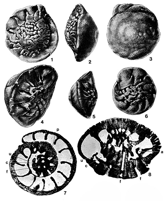
Description Loeblich, A. R., Tappan, H. N., 1987: Foraminiferal genera and their classification. Van Nostrand, Reinhold Co. New York 1728 pp. Plate 762, Figs. 1-8: M. Eocene (Lutetian), Blaye, Dept. Gironde, France. 1-3, Opposite sides and edge view of holotype, x 25; 4, oblique view of umbilical side, broken open to show basal intercameral foramen, areal foramen, and spiral canal opened at umbilical margin of chamber wall, x 25; 5, 6, edge and umbilical views of paratype with complete last chamber showing slitlike interiomarginal aperture, sutural chamber Iobes broken on last two chambers exposing part of septal passage beneath, x 25; 7, 8, oblique equatorial section, x 60, and axial section, x 64, showing areal foramen (a), spiral canal (c), funnel (f), septal passage (p), and umbilical flap (u), (from Parvati, 1971).
JPG file - 229.13 kB - 564 x 673 pixels
added on 2023-12-0436 viewsForaminifera taxaDigital photo, entire species Medocia blayensis Parvati, 1971 †checked Le Coze, François 2023-12-04
 Comment (0)
Disclaimer: Foraminifera does not exercise any editorial control over the information displayed here. However, if you come across any misidentifications, spelling mistakes or low quality pictures, your comments would be very much appreciated. You can reach us by emailing info@marinespecies.org, we will correct the information or remove the image from the website when necessary or in case of doubt.