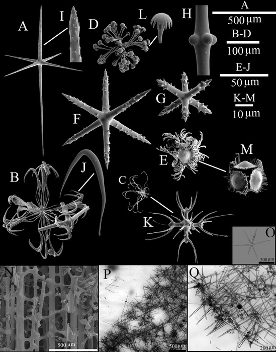
Spicules and skeleton
Description A, dermal or atrial hexactin; B, drepanocome I; C, drepanocome II; D, spirodiscohexaster; E, plumicome; F, microhexactin I; G, microhexactin II; H, detail of tubercles in the middle of a diactine; I, detail of the pinular ray of a hexactin; J, detail of the hook-like secondary ray of a drepanocome I; K, detail of the middle part of a drepanocome II; L, detail of the tooth disc of a spirodiscohexaster; M, detail of the whorl of a plumicome; N, spicules of the peduncle; O–Q, LM images of spicule and skeleton; O, choanosomal hexactin; P, tangential view of choanosomal structure; Q, transversal view of choanosomal structure.
JPG file - 969.94 kB - 2 008 x 2 562 pixels
Extra information
Original Width: 2008 pixels
Original Height: 2562 pixels
Orientation: 1: Horizontal (normal)
added on 2021-10-30204 viewsPorifera taxaDigital photo, part of species Saccocalyx microhexactin Gong, Li & Qiu, 2015checked Cárdenas, Paco 2021-10-30
Original Width: 2008 pixels
Original Height: 2562 pixels
Orientation: 1: Horizontal (normal)
 Comment (0)