Fukuyoa koreansis
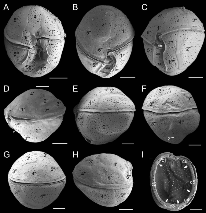
Description Scanning electron micrographs (SEM) of Fukuyoa koreansis sp. nov. (Strain LIMS-PS-2399). (A, B) Ventral view.(C) Ventral-left lateral view.
(D, E) Left lateral view.
(F, G) Dorsal-right lateral view.
(H) Right lateral view.
(I) Detail of the cingular plates. Small arrows indicate the five sutures separating the six cingular plates
(c1–c6). Scale bars: A–I = 10 μm. JPG file - 168.99 kB - 1 400 x 1 456 pixels added on 2023-03-15520 viewsTraits taxaMicroscope Fukuyoa koreensis Zhun Li, J.S.Park, N.S.Kang, K.-W.Lee & H.H. Shin, 2021checked Guiry, Michael D. 2023-03-15
 Comment (0)