Photogallery

P. bresslaui
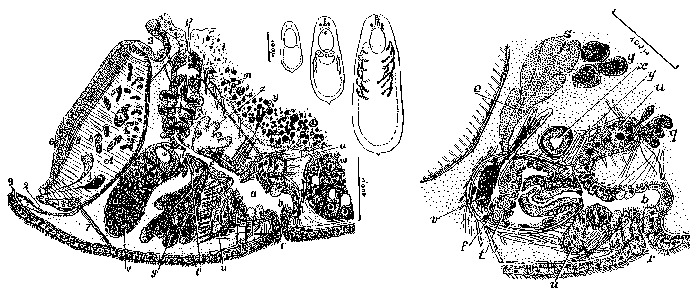
Description Fig. 81 (right): Sagittal section, seen from the left hand side, with the male apparatus (combined). e, efferent duct. f, diafragma. g, shell glands. q, pyriform glandular appendages. r, gonopore. s, granular secretion glands. t, vesicle with granular secretion. u, pyriform appendages. v, seminal vesicle. x, diverticle of the female genital canal y, sphincter of the female genital canal. Fig. 82 (middle): Freshly-hatched animal. Medio-adult individual, ventral view with mouth, gonopore and protonephridia; because of transparency an egg is visible. Adult worm, dorsal view, with the testicles not yet fully grown.
Fig. 83 (left). Sagittal section, seen from the right hand side, with the female apparatus (combined). a, atrium superior. b. atrium inferior. g, shell glands. l, glands at the opening of the oviduct. m, bursa. o, ovarium. p, bursa-intestinal pore. r, gonopore. u, pyriform appendages. w, vitellarium. y, sphincter of the female genital canal. z. single-nucleated, lobate glands.
GIF file - 60.95 kB - 1 477 x 627 pixels added on 2017-04-0774 viewsWoRMS taxaScan of photo Phaenocora bresslaui Marcus, 1946checked Tyler, Seth 2017-04-07
This work is licensed under a Creative Commons Attribution-NonCommercial-ShareAlike 4.0 International License
Click here to return to the thumbnails overview
