Photogallery

Kataplana celeretrix
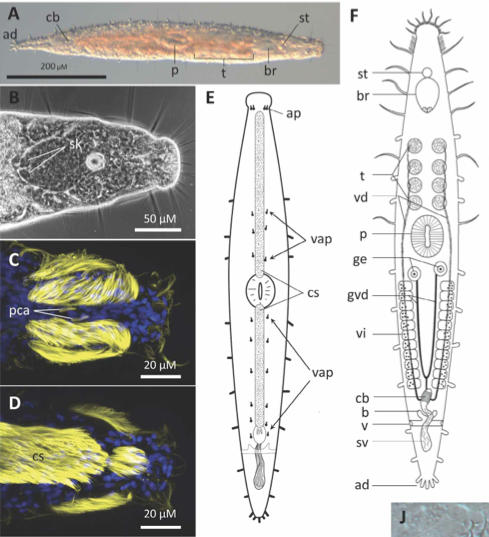
Description Anterior end is to the right in Figs A-D, H & I, and to the top in the remaining figures. A. Dorsal view of live animal, unsqueezed; ad, adhesive papillae of tailplate; br, brain; cb, copulatory bulb; p, pharynx; st, statocyst; t, testes. B. Phase-contrast micrograph of head region; paired Sehkolben (sk) are embedded in the posterior margin of the brain. C. CLSM stack of head, dorsal view; head ciliation is shown along with a pair of pericerebral ciliary aggregates (pca). D. CLSM stack of head, ventral view; head ciliation is shown along with the anterior part of the creeping sole (cs). E Schematic ventral view of animal, showing anterior pair of adhesive papillae (ap); ciliated creeping sole and ventral row of adhesive papillae (vap). F. Dorsal-view reconstruction of internal anatomy; b, bursa; ge, germaria; gvd, germo-vitelloducts; sv, seminal vesicle; v, paired vaginal openings; vd, vasa deferentia; vi, vitellaria.
PNG file - 240.66 kB - 489 x 537 pixels
added on 2017-04-07102 viewsWoRMS taxaScan of photo Kataplana celeretrix Bursey, Smith & Litviatis, 2012checked Tyler, Seth 2017-04-07
This work is licensed under a Creative Commons Attribution-NonCommercial-ShareAlike 4.0 International License
Click here to return to the thumbnails overview
