WoRMS Photogallery

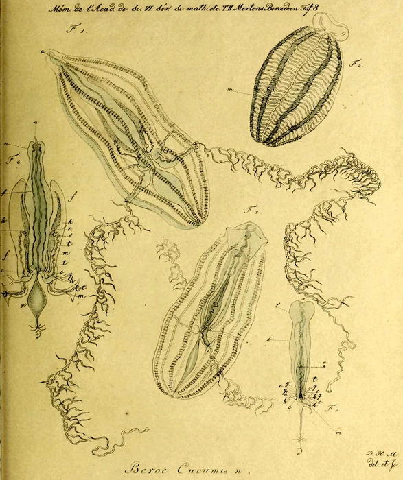
Hormiphora_cucumis_Holotype_as_Beroe_cucumis in Mertens (1833)
Description Beroë cucumis in mehreren Ansichten.
Fig. 1. Diesselbe im ausgedehnten Zustande mit zwei ausgebreiteten von ihrem Ursprunge an sichtbaren Fangfäden.
Fig. 2. Dasselbe Thier nur eine Fläche, worauf ein vollständiger, am Ende abgeschnittener Fangfaden sichtbar ist, dem Beschauer zuwendend.
Fig. 3. Das Thier contrahirt mit den in ihren Kanal, dessen Oeffnung m bezeichnet, zurückgezogenen Fangfäden.
Fig. 4. Die inneren Organe des Thiers. Daran a die Falte die über den Nahrungskanal läuft, n der Raum, in welchem der Nahrungskanal liegt, f die Leberschläuche, k die Säcke, worin die Eierstöcke liegen, e die Eierstöcke, s die neben der Mitte der Eierstöcke befindliche Fangfadenbasis.
t der Fangfaden, m der Kanal desselben, c der Anfang der arteriellen Gefässe, welehe Aeste g c, h zu den Rippen senden, r die Anschwellung des Darmes, d die Narbe.
Fig. 5. Dieselben Theile von der Seite, a der Mund, d die Narbe, l der Raum für den Verdauungskanal (k), e der Eierstock, s die Basis des Fangfadens, c der Stamm der zu den Rippen führenden Gefässe (gh), t der Fangfaden, m der Kanal desselben
Beroë cucumis in several views.
Fig. 1: The same animal in the expanded state with two spread out filaments [tentacles] visible from their origin.
Fig. 2: The same animal with only one surface, on which a complete thread [tentacle] cut off at the end is visible, facing the observer.
Fig. 3: The animal contracted with the threads [tentacles] retracted into their canals, the opening of which is marked by m. The internal organs of the animal.
Fig. 4 The internal organs of the animal. The following are shown: a the fold which runs over the feeding canal, n the space in which the feeding canal lies, f the liver tubes, k the sacs in which the ovaries lie, e the ovaries, s the base of the tentacle next to the middle of the ovaries.
t the tentacle, m the channel of the same, c the beginning of the arterial vessels, which send branches g c, h to the ribs [comb rows], r the swelling of the intestine, d the scar.
Fig. 5. the same parts from the side, a the mouth, d the stigma, l the space for the alimentary canal (k), e the ovary, s the base of the trap filament, c the trunk of the vessels leading to the ribs (gh), t the trap filament, m the canal of the same.
PNG file - 3.09 MB - 1 584 x 1 888 pixels
added on 2021-07-101 595 viewsWoRMS taxaScan of drawing Beroe cucumis Mertens, 1833 accepted as Hormiphora cucumis (Mertens, 1833)checked Lindsay, Dhugal 2021-07-10
Download full size
This work is licensed under a Creative Commons Attribution-NonCommercial-ShareAlike 4.0 International License
Click here to return to the thumbnails overview
 Comment (0)
 Click here to add a comment.
* indicates a required field.
Disclaimer: WoRMS does not exercise any editorial control over the information displayed here. However, if you come across any misidentifications, spelling mistakes or low quality pictures, your comments would be very much appreciated. You can reach us by emailing info@marinespecies.org or adding a comment, we will correct the information or remove the image from the website when necessary or in case of doubt.