WoRMS Photogallery

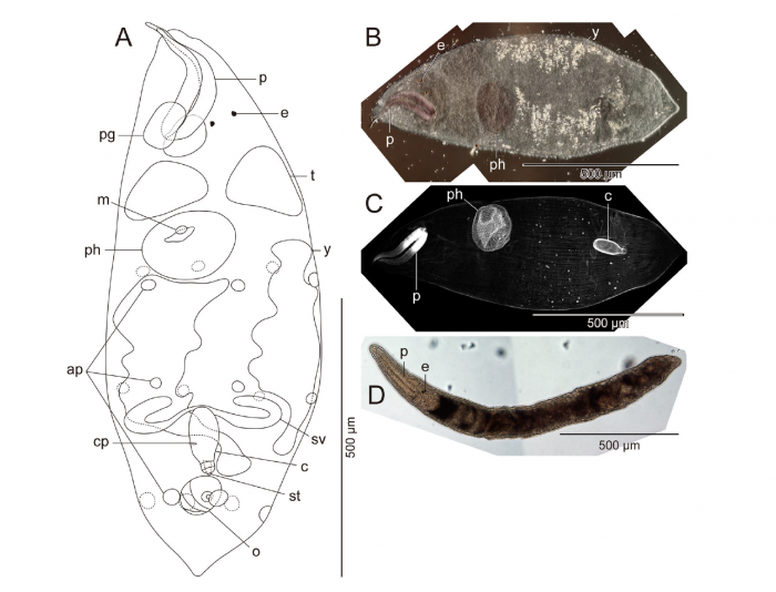
Proschizorhynchella magnoliae sp. nov
Description A. Illustration of fixed specimen showing arrangement of various internal organs, ICHUM 4859 (holotype); B. Photograph of fluorescent-staining specimen of the entire animal taken under a bright field, ICHUM 4859 (holotype); C. Confocal laser scanning micrograph, ICHUM 4859 (holotype); D. Composite photomicrograph taken in life, ICHUM 4849 (paratype). Abbreviations: ap, adhesive papilla; c, male copulatory organ; cp, common genital pore; e, eye; m, mouth; o, ovary; p, proboscis; pg, proboscis gland sac; ph, pharynx; st, stylet; sv, seminal vesicle; t, testis; y, yolk gland.
PNG file - 1.14 MB - 1 516 x 1 158 pixels
added on 2018-08-132 165 viewsWoRMS taxaScan of drawing Proschizorhynchella magnoliae Takeda & Kajihara, 2018checked Artois, Tom 2018-08-13
Download full size
This work is licensed under a Creative Commons Attribution-NonCommercial-ShareAlike 4.0 International License
Click here to return to the thumbnails overview
 Comment (0)
 Click here to add a comment.
* indicates a required field.
Disclaimer: WoRMS does not exercise any editorial control over the information displayed here. However, if you come across any misidentifications, spelling mistakes or low quality pictures, your comments would be very much appreciated. You can reach us by emailing info@marinespecies.org or adding a comment, we will correct the information or remove the image from the website when necessary or in case of doubt.