WoRMS Photogallery

Biarmifer nesiotes
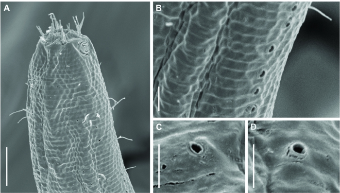
Description Biarmifer nesiotes sp. nov., scanning electron micrographs, male. (A) Anterior body region. (B) Anterior most lateral pore-like structures. (C) Detail of a pore complex in the precloacal region. (D) Detail of a lateral pore-like structure in the precloacal region. Scale bars: A = 20 µm; B-D = 5 µm.
JPG file - 1.81 MB - 1 503 x 853 pixels
Extra information
FileName: 156505.jpg
FileDateTime: 1677147372
FileSize: 1813192
FileType: 2
MimeType: image/jpeg
SectionsFound: ANY_TAG, IFD0, THUMBNAIL, EXIF
COMPUTED.html: width="1503" height="853"
COMPUTED.Height: 853
COMPUTED.Width: 1503
COMPUTED.IsColor: 0
COMPUTED.ByteOrderMotorola: 1
COMPUTED.Thumbnail.FileType: 2
COMPUTED.Thumbnail.MimeType: image/jpeg
ImageDescription: Biodiversity, Marine Biology, Taxonomy, Zoology
Orientation: 1
XResolution: 3000000/10000
YResolution: 3000000/10000
ResolutionUnit: 2
Software: Adobe Photoshop 23.1 (Windows)
DateTime: 2023:02:23 10:40:42
Exif_IFD_Pointer: 228
ColorSpace: 65535
ExifImageWidth: 1503
ExifImageLength: 853
added on 2023-02-231 588 viewsWoRMS taxaScan of photo Biarmifer nesiotes Cunha, Fonseca & Amaral, 2023checked Bezerra, Tânia Nara 2023-02-23
FileName: 156505.jpg
FileDateTime: 1677147372
FileSize: 1813192
FileType: 2
MimeType: image/jpeg
SectionsFound: ANY_TAG, IFD0, THUMBNAIL, EXIF
COMPUTED.html: width="1503" height="853"
COMPUTED.Height: 853
COMPUTED.Width: 1503
COMPUTED.IsColor: 0
COMPUTED.ByteOrderMotorola: 1
COMPUTED.Thumbnail.FileType: 2
COMPUTED.Thumbnail.MimeType: image/jpeg
ImageDescription: Biodiversity, Marine Biology, Taxonomy, Zoology
Orientation: 1
XResolution: 3000000/10000
YResolution: 3000000/10000
ResolutionUnit: 2
Software: Adobe Photoshop 23.1 (Windows)
DateTime: 2023:02:23 10:40:42
Exif_IFD_Pointer: 228
ColorSpace: 65535
ExifImageWidth: 1503
ExifImageLength: 853
This work is licensed under a Creative Commons Attribution-NonCommercial-ShareAlike 4.0 International License
Click here to return to the thumbnails overview
 Comment (0)
 Click here to add a comment.
* indicates a required field.
Disclaimer: WoRMS does not exercise any editorial control over the information displayed here. However, if you come across any misidentifications, spelling mistakes or low quality pictures, your comments would be very much appreciated. You can reach us by emailing info@marinespecies.org or adding a comment, we will correct the information or remove the image from the website when necessary or in case of doubt.