WoRMS Photogallery

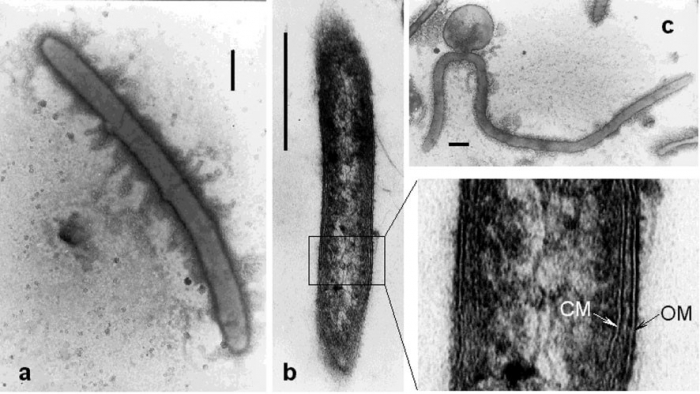
Caldithrix abyssi strain LF13T
Description (a) Electron micrograph of a negatively stained cell of strain LF13T. Bar, 0.5 mm.(b) Electron micrograph of thinsection of a cell of strain LF13T, exhibiting cell-wall structure. CM, cytoplasmic membrane; OM, outer membrane. Bar, 0.5 mm.
(c) Electron micrograph of thin-section of a cell of strain LF13T, exhibiting formation of a spherical body. Bar, 0.5 mm.
Source: https://doi.org/10.1099/ijs.0.02390-0
JPG file - 120.43 kB - 886 x 499 pixels added on 2018-06-291 964 viewsWoRMS taxaMicroscope Caldithrix abyssi Miroshnichenko et al., 2003
This work is licensed under a Creative Commons Attribution-NonCommercial-ShareAlike 4.0 International License
Click here to return to the thumbnails overview
 Comment (0)
 Click here to add a comment.
* indicates a required field.
Disclaimer: WoRMS does not exercise any editorial control over the information displayed here. However, if you come across any misidentifications, spelling mistakes or low quality pictures, your comments would be very much appreciated. You can reach us by emailing info@marinespecies.org or adding a comment, we will correct the information or remove the image from the website when necessary or in case of doubt.