WoRMS taxon details
Tetranemertes bifrost Cherneva, Ellison, Zattara, Norenburg, Schwartz, Junoy & Maslakova, 2023
1739517 (urn:lsid:marinespecies.org:taxname:1739517)
accepted
Species
marine, brackish, fresh, terrestrial
recent only
Cherneva, I.; Ellison, C. I.; Zattara, E. E.; Norenburg, J. L.; Schwartz, M. L.; Junoy, J.; Maslakova, S. A. (2023). Seven new species of Tetranemertes Chernyshev, 1992 (Monostilifera, Hoplonemertea, Nemertea) from the Caribbean Sea, western Pacific, and Arabian Sea, and revision of the genus. <em>ZooKeys.</em> 1181: 167-200., available online at https://doi.org/10.3897/zookeys.1181.109521 [details] 
Holotype USNM 1618678
Holotype USNM 1618678 [details]
Description External appearance of live specimens. Long, thin, thread-like body. Can stretch much more than 200 mm long, and up to 0.5...
Distribution Caribbean Sea: Bocas del Toro, Panamá, also common at Fuerte Sherman, Colon, Panamá; photographed off Puerto Rico, draped...
Etymology The name refers to the bright, colorful iridescent stripes and spots characterizing this species. Bifrost, the rainbow...
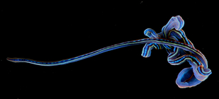
Description External appearance of live specimens. Long, thin, thread-like body. Can stretch much more than 200 mm long, and up to 0.5 mm wide at the head; body up to 0.7 mm wide. Body slightly compressed dorso-laterally in cross-section throughout most of its length. The caudal end is rounded with no obvious modifications. Tetranemertes bifrost sp. nov. is, perhaps, the most spectacularly colored nemertean in the Caribbean, if not the world (Fig. 4A–F). Larger individuals present a black to deep purple body dorsally, with a discontinuous bright blue mid-dorsal longitudinal stripe running from anterior to posterior tip of body. The blue stripe is flanked by two continuous dorso-lateral stripes that tend to appear orange to yellow in larger individuals, or sky blue to luminescent green in smaller individuals. The flanking stripes begin at the level of the cerebral organ furrow, and continue to the posterior tip of body. The pigment composing the stripes is highly iridescent, so that perceived coloration changes with type and direction of incident light. Ventral side is typically bright blue, pigment
scattered over the dark background in a form of numerous tiny speckles (Fig. 4A, D, F). When placed in a dish, worms tangle into a writhing mass, and secrete transparent sticky mucus when handled. Move by ciliary gliding, often with a peristaltic wave passing along the anterior part of the body. When mechanically perturbed, contract into a loose coil. The anterior head region is less contractile, causing an obvious discontinuity in width. Blood is colorless. Remarkably resistant to breaking while being extracted from complex substratum.
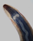
The head has a characteristic, narrow diamond or spearhead shape, vaguely reminiscent of a viper’s head, its anterior region demarcated from the rest of body both by width and a single ventral cephalic furrow, formed by the midventral fusion of the cerebral organ furrows. The cephalic furrow is located anterior to the cerebral ganglia, runs from the lateral sides toward the ventral midline forming a shallow forward-pointing “V”. Margins of the head are clear, translucent; the anterior-most mid-dorsal part of the head often reddish brown (Fig. 4G). Small
yellow-orange iridescent speckles are scattered on the lateral sides of the head at the level of the cephalic furrow (Fig. 4E). Mouth and rhynchopore combine into a single ventral anterior rhynchostomopore. Posterior cephalic furrows lacking. Numerous small reddish brown ocelli (10–20 on each side of head) are arranged in four longitudinal rows (Fig. 4E–G). The medial rows, which are not visible in ventral view, reach as far back as anterior margin of cerebral ganglia. The lateral rows have fewer eyes, reach slightly past the cephalic furrow and are visible in ventral view (Fig. 4F). Ocelli are somewhat obscured by the dark pigment of the head, and best seen in worms compressed between a slide and a cover-glass (Fig. 4G). When viewed from dorsal side appear to form two rows (because the two rows on each side of head are almost directly on top of each other). Cerebral organs are small and inconspicuous, difficult to see in living specimens, even when compressed under a glass slide and viewed under a compound microscope. Anterior to the cephalic furrow, the head is dorso-ventrally flattened; posterior to it, there is a dorsal bulge over the brain, and the body is rounder in cross-section than the anterior tip. The anterior margin of the head is bluntly pointed, indented anteriorly by the rhynchostomopore. The cerebral ganglia are clear, and are located at the base of the “spearhead”, connected by fairly wide cerebral commissures (Fig. 4G).
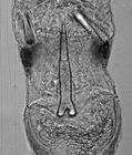
Rhynchocoel and proboscis. Rhynchocoel does not exceed 1/3 of body length. Proboscis transparent, with two distinctive regions separated by a stylet region. The anterior proboscis is much shorter than the posterior, and the stylet bulb has a length to width ratio of ~ 1. The proboscis is arm [details]
Distribution Caribbean Sea: Bocas del Toro, Panamá, also common at Fuerte Sherman, Colon, Panamá; photographed off Puerto Rico, draped...
Distribution Caribbean Sea: Bocas del Toro, Panamá, also common at Fuerte Sherman, Colon, Panamá; photographed off Puerto Rico, draped over an unknown fan coral (in 1970s by Smithsonian photographer Kjell Sandved). [details]
Etymology The name refers to the bright, colorful iridescent stripes and spots characterizing this species. Bifrost, the rainbow...
Etymology The name refers to the bright, colorful iridescent stripes and spots characterizing this species. Bifrost, the rainbow bridge in the Norse mythology, reaches between Midgard, the human Earth, and Asgard, the realm of the gods. Some authors state that the name Bifrost means “shimmering path” or “the swaying road to heaven”, and that it might be inspired by the Milky Way. [details]
Norenburg, J.; Gibson, R.; Herrera Bachiller, A.; Strand, M. (2024). World Nemertea Database. Tetranemertes bifrost Cherneva, Ellison, Zattara, Norenburg, Schwartz, Junoy & Maslakova, 2023. Accessed through: World Register of Marine Species at: https://www.marinespecies.org/aphia.php?p=taxdetails&id=1739517 on 2024-05-09
![]() The webpage text is licensed under a Creative Commons Attribution 4.0 License
The webpage text is licensed under a Creative Commons Attribution 4.0 License
original description
Cherneva, I.; Ellison, C. I.; Zattara, E. E.; Norenburg, J. L.; Schwartz, M. L.; Junoy, J.; Maslakova, S. A. (2023). Seven new species of Tetranemertes Chernyshev, 1992 (Monostilifera, Hoplonemertea, Nemertea) from the Caribbean Sea, western Pacific, and Arabian Sea, and revision of the genus. <em>ZooKeys.</em> 1181: 167-200., available online at https://doi.org/10.3897/zookeys.1181.109521 [details] 
From editor or global species database
Description External appearance of live specimens. Long, thin, thread-like body. Can stretch much more than 200 mm long, and up to 0.5 mm wide at the head; body up to 0.7 mm wide. Body slightly compressed dorso-laterally in cross-section throughout most of its length. The caudal end is rounded with no obvious modifications. Tetranemertes bifrost sp. nov. is, perhaps, the most spectacularly colored nemertean in the Caribbean, if not the world (Fig. 4A–F). Larger individuals present a black to deep purple body dorsally, with a discontinuous bright blue mid-dorsal longitudinal stripe running from anterior to posterior tip of body. The blue stripe is flanked by two continuous dorso-lateral stripes that tend to appear orange to yellow in larger individuals, or sky blue to luminescent green in smaller individuals. The flanking stripes begin at the level of the cerebral organ furrow, and continue to the posterior tip of body. The pigment composing the stripes is highly iridescent, so that perceived coloration changes with type and direction of incident light. Ventral side is typically bright blue, pigmentscattered over the dark background in a form of numerous tiny speckles (Fig. 4A, D, F). When placed in a dish, worms tangle into a writhing mass, and secrete transparent sticky mucus when handled. Move by ciliary gliding, often with a peristaltic wave passing along the anterior part of the body. When mechanically perturbed, contract into a loose coil. The anterior head region is less contractile, causing an obvious discontinuity in width. Blood is colorless. Remarkably resistant to breaking while being extracted from complex substratum.
The head has a characteristic, narrow diamond or spearhead shape, vaguely reminiscent of a viper’s head, its anterior region demarcated from the rest of body both by width and a single ventral cephalic furrow, formed by the midventral fusion of the cerebral organ furrows. The cephalic furrow is located anterior to the cerebral ganglia, runs from the lateral sides toward the ventral midline forming a shallow forward-pointing “V”. Margins of the head are clear, translucent; the anterior-most mid-dorsal part of the head often reddish brown (Fig. 4G). Small
yellow-orange iridescent speckles are scattered on the lateral sides of the head at the level of the cephalic furrow (Fig. 4E). Mouth and rhynchopore combine into a single ventral anterior rhynchostomopore. Posterior cephalic furrows lacking. Numerous small reddish brown ocelli (10–20 on each side of head) are arranged in four longitudinal rows (Fig. 4E–G). The medial rows, which are not visible in ventral view, reach as far back as anterior margin of cerebral ganglia. The lateral rows have fewer eyes, reach slightly past the cephalic furrow and are visible in ventral view (Fig. 4F). Ocelli are somewhat obscured by the dark pigment of the head, and best seen in worms compressed between a slide and a cover-glass (Fig. 4G). When viewed from dorsal side appear to form two rows (because the two rows on each side of head are almost directly on top of each other). Cerebral organs are small and inconspicuous, difficult to see in living specimens, even when compressed under a glass slide and viewed under a compound microscope. Anterior to the cephalic furrow, the head is dorso-ventrally flattened; posterior to it, there is a dorsal bulge over the brain, and the body is rounder in cross-section than the anterior tip. The anterior margin of the head is bluntly pointed, indented anteriorly by the rhynchostomopore. The cerebral ganglia are clear, and are located at the base of the “spearhead”, connected by fairly wide cerebral commissures (Fig. 4G).
Rhynchocoel and proboscis. Rhynchocoel does not exceed 1/3 of body length. Proboscis transparent, with two distinctive regions separated by a stylet region. The anterior proboscis is much shorter than the posterior, and the stylet bulb has a length to width ratio of ~ 1. The proboscis is arm [details]
Diagnosis Tetranemertes bifrost sp. nov. differs from all other species of this genus by its distinctive color: purple to black dorsally, with a pattern of bright iridescent longitudinal stripes and spots, which may appear blue, green, yellow and orange, depending on the type of lighting, background, and individual, and blue ventrally. Additionally, it differs from T. antonina, T. hermaphroditica, and T. paulayi sp. nov. by basis of central stylet posteriorly forked, and from the first two species by having stylets sculpted with spiral groves. At the molecular level, COI barcodes of sequenced specimens clearly differentiate it from other species of Tetranemertes (Table 5). [details]
Distribution Caribbean Sea: Bocas del Toro, Panamá, also common at Fuerte Sherman, Colon, Panamá; photographed off Puerto Rico, draped over an unknown fan coral (in 1970s by Smithsonian photographer Kjell Sandved). [details]
Etymology The name refers to the bright, colorful iridescent stripes and spots characterizing this species. Bifrost, the rainbow bridge in the Norse mythology, reaches between Midgard, the human Earth, and Asgard, the realm of the gods. Some authors state that the name Bifrost means “shimmering path” or “the swaying road to heaven”, and that it might be inspired by the Milky Way. [details]
Habitat Free living, benthic marine worms inhabiting coral rubble, gravel, and shell hash. Often found stretched between nooks and crannies of the substratum. [details]
Reproduction Oocytes, measuring 50–80 micron in diameter were noted in specimen CB057_18_03 collected in August 2018. [details]


