Integrated Marine Information System (IMIS)
Persons | Institutes | Publications | Projects | Datasets
Haplogonaria schillingi
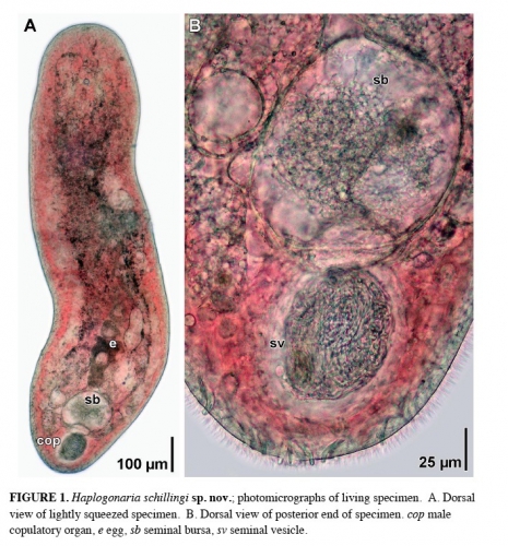
Description photomicrographs of living specimen. A. Dorsal view of lightly squeezed specimen. B. Dorsal view of posterior end of specimen. cop male copulatory organ, e egg, rg rhabdoid gland, sb seminal bursa, sv seminal vesicle.
JPG file - 240.32 kB - 707 x 761 pixels
Extra information
FileName: 124572.jpg
FileDateTime: 1447796772
FileSize: 240320
FileType: 2
MimeType: image/jpeg
SectionsFound: ANY_TAG, IFD0, EXIF
COMPUTED.html: width="707" height="761"
COMPUTED.Height: 761
COMPUTED.Width: 707
COMPUTED.IsColor: 1
COMPUTED.ByteOrderMotorola: 1
Orientation: 1
Exif_IFD_Pointer: 38
ExifImageWidth: 707
ExifImageLength: 761
added on 2017-04-072 119 views
FileName: 124572.jpg
FileDateTime: 1447796772
FileSize: 240320
FileType: 2
MimeType: image/jpeg
SectionsFound: ANY_TAG, IFD0, EXIF
COMPUTED.html: width="707" height="761"
COMPUTED.Height: 761
COMPUTED.Width: 707
COMPUTED.IsColor: 1
COMPUTED.ByteOrderMotorola: 1
Orientation: 1
Exif_IFD_Pointer: 38
ExifImageWidth: 707
ExifImageLength: 761
This work is licensed under a Creative Commons Attribution-NonCommercial-ShareAlike 4.0 International License
Click here to return to the thumbnails overview